Back Bones Diagram / The Vertebral Column Joints Vertebrae Vertebral Structure
Back Bones Diagram / The Vertebral Column Joints Vertebrae Vertebral Structure. The human body is an incredible machine. Spinal anatomy is a remarkable combination of strong bones, flexible ligaments and tendons, large muscles and highly sensitive nerves. All the images are in vector format, allowing an optimal web display with zoom and shifting of the anatomical images. The vertebrae, which stack like spools of thread, support the back and protect the spinal cord. A tough, springy disc of cartilage sits between the vertebrae of your spine.
ads/bitcoin1.txt
In the back and elsewhere in the body, tendons attach muscles to bones. It is designed to be incredibly strong, protecting the highly sensitive nerve roots, yet highly flexible, providing for mobility on many different planes. 12 photos of the human back bones diagram. You can read more detail about these important bones in the arm from the following description and diagram. It is like that for several reasons, all of which you can understand by looking at the anatomy of the thoracic spine.
Low Back Pain Wikipedia from upload.wikimedia.org The vertebral column is a series of approximately 33 bones called vertebrae, which are separated by intervertebral discs. The rib cage also anchors the bones of the head, neck, shoulders, and arms to the trunk of the body. It is like that for several reasons, all of which you can understand by looking at the anatomy of the thoracic spine. Lower jaw (mandible) collar bone. The lumbar spine connects to the thoracic spine above and the hips below. Over 3000+ pages with full illustrations and diagrams. It consists of 5 lumbar vertebra that are numbered 1 through 5 from top to bottom i.e. Here we will attempt to provide a brief overview of lumbar spinal anatomy.
Lateral labeled diagram of the human vertebral spinal column showing vertebrae numbering order and the 5 different regions of the spine.
ads/bitcoin2.txt
Female pelvis bones images 12 photos of the female pelvis bones images female human pelvis images, female pelvic bones images, bone, female human pelvis. Powerful muscles that move the head and arms attach to these bones as well. You can read more detail about these important bones in the arm from the following description and diagram. It is like that for several reasons, all of which you can understand by looking at the anatomy of the thoracic spine. This diagram determines the possible causes of a specific event or problem. The spine diagram the spine diagram shown below, consists of many bones or vertebrae,soft discs,the spinal cord, and spinal nerves. A tough, springy disc of cartilage sits between the vertebrae of your spine. Bone diagram back skeletal dysplasias affect the development and growth of cartilage bones and joints causing abnormally shaped bones especially in the head spine and f i g u r e 1 diagram of. But, they are common in the back and can cause pain. Bones of the body blank diagram, bones of the body jingle, bones of the body quiz game, number of bones of the body, bone, bones of the body anatomy, bones of the body blank. Lateral labeled diagram of the human vertebral spinal column showing vertebrae numbering order and the 5 different regions of the spine. Bones of the chest and upper back / the muscles, bones, ligaments, and tendons in the back can all be injured and cause back. The first seven bones (vertebrae) of your spine form your neck.
Back bones diagram, human back bones skeleton, human back muscles and bones, human backbone structure, pictures of human back bones, bone. The vertebral column is a series of approximately 33 bones called vertebrae, which are separated by intervertebral discs. Your lower back contains 5 vertebral bones stacked above each other with intervertebral discs in between. Can you feel the bumps of your vertebrae along your back? It is the surface of the body opposite from the chest.
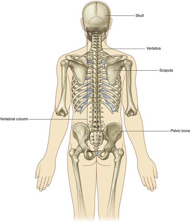
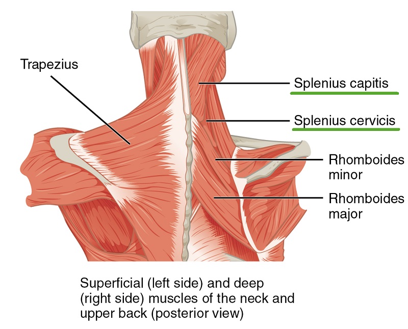
Back Clinical Gate from clinicalgate.com The column can be divided into five different regions, with each region characterised by a different vertebral structure. The bones of the pelvis and lower back work together to support the body's weight, anchor the abdominal and hip muscles, and protect the delicate vital organs of the vertebral and abdominopelvic cavities. All the images are in vector format, allowing an optimal web display with zoom and shifting of the anatomical images. Your lower back contains 5 vertebral bones stacked above each other with intervertebral discs in between. The bones of the chest and upper back combine to form the strong, protective rib cage around the vital thoracic organs such as the heart and lungs. The vertebral column of the lower back includes the five lumbar vertebrae, the sacrum, and the coccyx. The red lines point individual bones and the names are writen in singular, the blue lines conect to group of bones and are in plural form. There are three parts to the trapezius.
Bones of the body blank diagram, bones of the body jingle, bones of the body quiz game, number of bones of the body, bone, bones of the body anatomy, bones of the body blank.
ads/bitcoin2.txt
Anatomical diagrams of the spine and back. Here we will attempt to provide a brief overview of lumbar spinal anatomy. Spinal anatomy is a remarkable combination of strong bones, flexible ligaments and tendons, large muscles and highly sensitive nerves. But, they are common in the back and can cause pain. Diagram of a human female skeleton, back view. Lateral labeled diagram of the human vertebral spinal column showing vertebrae numbering order and the 5 different regions of the spine. L1, l2, l3, l4, and l5. The human body is an incredible machine. Related posts of human back bones diagram female pelvis bones images. The vertebral column is a series of approximately 33 bones called vertebrae, which are separated by intervertebral discs. File human arm bones diagram svg wikipedia. Lower jaw (mandible) collar bone. It consists of 5 lumbar vertebra that are numbered 1 through 5 from top to bottom i.e.
The lower part of the trapezius ascends and depresses the scapula, while the transverse or middle region of the trapezius is what retracts the. Individual anatomical structures include 2: Muscle or tendon injuries can occur anywhere in the body. The bones of the pelvis and lower back work together to support the body's weight, anchor the abdominal and hip muscles, and protect the delicate vital organs of the vertebral and abdominopelvic cavities. Bones, discs, and joints in your lower back.
Intrinsic Back Muscles Anatomy Of The Torso Medical Library from d3uigcfkiiww0g.cloudfront.net It is designed to be incredibly strong, protecting the highly sensitive nerve roots, yet highly flexible, providing for mobility on many different planes. Individual anatomical structures include 2: It is the surface of the body opposite from the chest. The neck (cervical) and low back (lumbar) regions have a slight concave curve, and the thoracic and sacral regions have a gentle convex curve (fig. File human arm bones diagram svg wikipedia. Lower jaw (mandible) collar bone. Use our interactive diagram to explore the different parts of the skeletal system. Related posts of human back bones diagram female pelvis bones images.
Divisions of the back bones.
ads/bitcoin2.txt
The neck (cervical) and low back (lumbar) regions have a slight concave curve, and the thoracic and sacral regions have a gentle convex curve (fig. Posted in bones , diagrams | tagged body skeleton , human skeletal anatomy , human skeleton , human skeleton anatomy , skeletal , skeletal anatomy , skeletal images. Bones of the pelvis and lower back. But, they are common in the back and can cause pain. Spinal vertebrae bone spine vertebra toracica spinal cord spine structure back diagram spine sections spinal cord vertebrae spinal structure health diagram. At the back of each bone in the spine (vertebra) are bony points called processes, which muscles attach to. Divisions of the back bones. There are three parts to the trapezius. The red lines point individual bones and the names are writen in singular, the blue lines conect to group of bones and are in plural form. The vertebrae, which stack like spools of thread, support the back and protect the spinal cord. Diagram of a human female skeleton, back view. Use our interactive diagram to explore the different parts of the skeletal system. It is the surface of the body opposite from the chest.
ads/bitcoin3.txt
ads/bitcoin4.txt
ads/bitcoin5.txt
0 Response to "Back Bones Diagram / The Vertebral Column Joints Vertebrae Vertebral Structure"


0 Response to "Back Bones Diagram / The Vertebral Column Joints Vertebrae Vertebral Structure"
Posting Komentar